

Prenatal Ultrasound in Warangal
Also called: Sonogram, prenatal ultrasound, fetal ultrasound
- SAMPLE
- Not required
- Report
- Same or next day
- PREPARATIONS
- No special preparation
The Scan at a glance
- What it checks
- U/S PREG test is a clinically significant diagnostic investigation used to evaluate, screen, or monitor underlying health conditions. It provides actionable insights that support early diagnosis, treatment planning, and long-term disease management.
- Report delivery
- For report delivery timelines, please contact the front desk or call +91 93901 10089.
- Test for
- Women, Family
- Age group
- 18+
- Commonly advised for
- Bleeding, pelvic pain, high-risk pregnancy issues
- Home collection
- No
What is a Complete Blood Count?
Know More About This Test
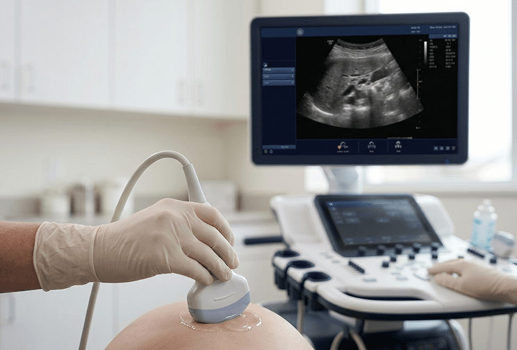
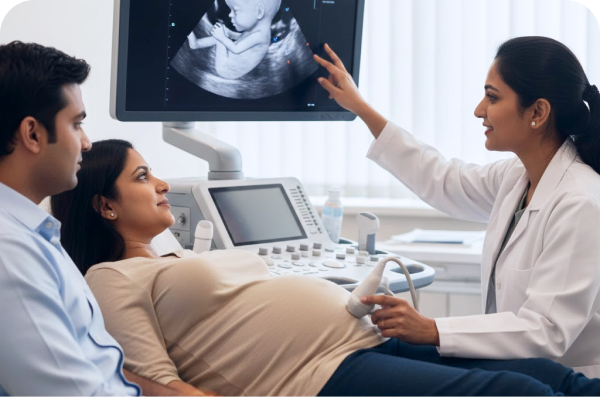
The Ultrasound Pregnancy test is a non-invasive imaging procedure that uses sound waves to visualize the developing fetus inside the uterus. It confirms pregnancy, estimates gestational age, assesses fetal viability, and detects abnormalities. It is a cornerstone of antenatal care and ensures maternal and fetal well-being.
Why Is This Scan Recommended?
This scan may be recommended to:
- Screen for structural abnormalities in the baby's brain, spine, heart, face, kidneys, limbs, and abdominal organs as part of routine mid-pregnancy assessment
- Confirm or reassess gestational age and estimated date of delivery, particularly if the first trimester dating scan was not performed
- Identify the location of the placenta and assess amniotic fluid volume - both of which are critical indicators of pregnancy health and safe delivery planning
- Detect soft markers of chromosomal abnormality such as thickened nuchal fold, absent nasal bone, or mild ventricular dilatation that may prompt further genetic investigation
- Monitor fetal growth in women with high-risk pregnancies, including those with diabetes, hypertension, or a history of pregnancy complications
- Assess cervical length at various stages of pregnancy to monitor the risk of preterm labor when clinically indicated
- Evaluate twin or multiple pregnancies and monitor for complications unique to multiple gestations
Symptoms or Conditions That May Require This Scan
This scan is recommended for all pregnant women beyond 16 weeks as part of routine antenatal care. It is particularly important in the following situations:
- Advanced maternal age (35 years or older), which carries a higher risk of chromosomal abnormalities such as Down syndrome
- A positive first-trimester screening result (nuchal translucency, NIPT, or combined screening) that requires anatomical follow-up
- A family history of congenital abnormalities, genetic syndromes, or structural defects in a previous child
- Reduced fetal movements or a fundal height measurement inconsistent with gestational age
- Vaginal bleeding, abdominal pain, or other symptoms that require assessment of the placenta and amniotic fluid
- Pregnancies conceived through IVF or assisted reproductive technologies, which are monitored more closely for fetal growth and structural development
- Medical conditions in the mother such as gestational diabetes, thyroid disorders, or autoimmune diseases that may affect fetal development
Advantages of the Pregnancy Ultrasound Scan (Above 16 Weeks)
The mid-pregnancy ultrasound is one of the most comprehensive assessments available during antenatal care, evaluating multiple organ systems, the placenta, and the amniotic environment in a single, non-invasive procedure. Advanced techniques such as 3D and 4D ultrasounds provide clearer, more detailed images, making it easier to identify complex conditions when required. Unlike blood-based screening tests that provide probability estimates, the anomaly scan offers direct visual evidence of the baby's structural development.
The detection rate of the screening second-trimester ultrasound scan is good in high-risk patients examined by a well-trained sonographer. The scan can also be sensitive in anomaly detection in low-risk women when a systematic searching pattern is followed. Its safety profile is well-established ultrasound has been used for over 40 years to monitor pregnancies, and the evidence has been reassuring that it is safe for both mother and baby. What Does the Scan Assess?
The pregnancy ultrasound scan beyond 16 weeks provides a structured, organ-by-organ assessment of the developing baby alongside an evaluation of the pregnancy environment:
- Brain and spine: Assessment for neural tube defects, hydrocephalus, and structural brain abnormalities
- Face: Evaluation for cleft lip and palate, and facial structural development
- Heart: Screening for congenital heart defects - a four-chamber view and outflow tract views are examined. Fetal echocardiography may be arranged separately for a more detailed cardiac assessment where indicated
- Chest: Assessment of lung development and detection of diaphragmatic hernia
- Abdominal wall and organs: Examination of the stomach, liver, bowel, kidneys, and bladder for structural and functional abnormalities
- Limbs and skeleton: Assessment of all four limbs for length, structure, and movement
- Placenta: Identification of position, maturity, and signs of abnormal implantation
- Amniotic fluid: Assessment of volume as a key indicator of fetal wellbeing and kidney function
- Fetal growth biometry: Measurements of head circumference, abdominal circumference, and femur length to calculate estimated fetal weight and confirm appropriate growth for gestational age
What Do the Results Mean?
- Normal findings: All assessed structures appear within expected parameters for the gestational age. The baby is growing appropriately and no significant structural abnormalities are identified. Routine antenatal care continues as planned
- Soft markers detected: Certain ultrasound findings - such as a thickened nuchal fold, absent nasal bone, or echogenic bowel - are associated with a mildly increased risk of chromosomal abnormality. These do not confirm a diagnosis but may prompt referral for genetic counselling or further testing
- Structural abnormality detected: A definitive structural finding such as a neural tube defect, cardiac anomaly, or renal malformation is identified. Further specialist evaluation, additional imaging, or invasive diagnostic testing may follow
- Incomplete scan: Even if the scan is normal, there is a small chance that the baby may still have a problem. Some conditions, including certain heart defects and bowel obstructions, may not be visible until later in the pregnancy. A repeat or follow-up scan may be recommended
- Growth concerns: Measurements significantly below expected ranges for gestational age may indicate intrauterine growth restriction, requiring closer monitoring and possible early delivery planning
Lifestyle Tips for a Healthy Pregnancy
- Attend all scheduled antenatal appointments and scans without delay - each scan is timed to coincide with a specific stage of development and serves a distinct clinical purpose
- Maintain a balanced, nutrient-rich diet adequate in folic acid, iron, calcium, and omega-3 fatty acids to support the baby's organ development during the second trimester
- Stay well hydrated throughout pregnancy, as adequate amniotic fluid volume is essential for the baby's development and for obtaining good scan images
- Avoid smoking, alcohol, and self-medication entirely during pregnancy, as these have well-documented adverse effects on fetal growth and structural development
- Manage chronic conditions such as gestational diabetes or hypertension carefully under medical supervision, as uncontrolled maternal health conditions directly affect fetal growth and development
- Report any change in fetal movement patterns, vaginal bleeding, or unusual abdominal symptoms to your obstetrician promptly, without waiting for the next scheduled appointment
Frequently Asked Questions (FAQs)
- Is fasting required before this scan? No - fasting is not required. Patients are generally advised to have a reasonably full bladder before the appointment to improve image quality.
- How long does the scan take? The mid-pregnancy anomaly scan typically takes approximately 30 minutes. A more detailed level II scan or fetal echocardiogram may take up to 45–60 minutes.
- Is the scan safe for the baby? Yes - ultrasound has been used for over 40 years to monitor pregnancies and the evidence has been reassuring that it is safe for both mother and baby.
- Can the scan detect Down syndrome? This scan can identify 50–60% of cases of Down syndrome, but first-trimester screening is more effective. Because 30–50% of Down syndrome cases appear normal on ultrasound, only an amniocentesis can provide a definitive chromosomal answer.
- What if the scan is inconclusive? If the baby's position does not allow complete examination of all structures, a repeat scan will be scheduled. At some centres, a free rescan is offered in such circumstances.
- How many ultrasound scans are recommended during pregnancy? The standard recommended schedule includes a first-trimester dating scan (11–13 weeks), a mid-trimester anomaly scan (18–22 weeks), and a third-trimester growth scan (28–32 weeks). Additional scans may be recommended based on individual clinical circumstances and risk factors.
Who should take this test
A pregnancy ultrasound checks the baby's growth and wellbeing using sound waves. It is advised if:
- Your doctor has scheduled a routine pregnancy scan
- You have bleeding or pelvic pain
- Your pregnancy is being monitored as high risk
- Your doctor wants to check the baby's growth and position
- Baby movements have reduced
Prenatal Ultrasound in Warangal
Worried about your Prenatal Ultrasound scan? Tapadia Diagnostic Centre in Warangal offers AERB-licensed Prenatal Ultrasound. Sonogram, prenatal ultrasound, fetal ultrasound is commonly prescribed for Bleeding, pelvic pain, high-risk pregnancy issues and suitable for 18+ Women, Family. Pay at the centre with no advance payment.
Frequently asked questions
Related Scans in Warangal



Centers You Can Choose To Visit
Vijayawada
- H.no 29-27-22, Suryanarayana street, suryaraopet vijayawada-520002
- Services: Lab
- Branch Incharge: Mr. Akhil : +91 6300387321
Get Direction
KPHB COLONY
- MIG H1, Plot No 273, above Visakhapatnam Coop Bank, Kukatpally Housing Board Colony, K P H B Phase 1, Kukatpally, Hyderabad, Telangana 500072
- Richards-9866362080
- Services - Lab, Digital X-Ray. ECG
Get Direction
Tapadia Diagnostic Center Bengaluru
- Sahakarnagar Main Rd, G Block, Sahakar Nagar, Byatarayanapura, Bengaluru, Karnataka 560092
- All facilities Available
- Branch Incharge: Mohammad Sarfaraz :+91 79896 92437
Get Direction
TAPADIA DIAGNOSTIC CENTRE (Main Branch)
- Main Branch
- 1-7-1072/A, RTC X Roads, Opp Saptagiri Theatre, Hyderabad, Telangana, India 500020.
- All facilities Available
- Give call at -8305555999
- Alternate Number -9390110098
Get Direction
SECUNDERABAD
- St. Thomas Spg Church Telugu Pastorate, near Sitara Hotel, Complex, Palika Bazar, Monda Market, Shivaji Nagar, Secunderabad, Telangana 500003
- Services: Lab, ECG
- Ravi-9100577378
Get Direction
Chhatrapati Sambhajinagar (Aurangabad)
- Bansilal Nagar, Station Road, Chhatrapati Sambhajinagar (Aurangabad).
- Services: MRI, CT Scan, Ultrasound, Lab, X Ray, ECG
- Amar Mundada: 94055 85858
Get Direction
Prenatal Ultrasound in Warangal
Rated 4.6 on Google From 6,477 patient reviews across our Hyderabad centres.
Every report, kept for you Past reports stay in your account and can be downloaded again.
NABL accredited Every result reviewed by a qualified pathologist before it reaches you.
Four decades of practice A family-run diagnostic centre, trusted by doctors and households since 1989
Medically Reviewed by Qualified Biochemists

Prenatal Ultrasound in Other Cities
MRI Knee - Both Joints in WarangalMRI Knee - Left Joint in WarangalMRI Knee - Right Joint in WarangalMRI C Spine in WarangalMRI LS Spine in WarangalMRI Brain in WarangalPulmonary Function Test in WarangalMRI - Brain/Spine/Knee in WarangalElectrocardiogram (ECG) in WarangalMammogram in WarangalChest X-ray in WarangalAbdominal Ultrasound in Warangal2D Echo in WarangalExercise Stress Test in WarangalPrenatal Ultrasound in WarangalX-Ray KUB in WarangalChest CT Scan in WarangalEEG in WarangalCT Head in WarangalHRCT Chest in WarangalAnomaly Scan in Warangal

 Made with Levo